The role of artificial intelligence in the diagnosis of liver lesions
Focal liver lesions (FLLs) can be difficult to detect, classify, and differentiate – especially when it comes to identifying which ones are benign and which are potentially malignant or may become so in the future. That’s where artificial intelligence (AI) can help reform the process. In our latest review, written in collaboration with Hospital de la Santa Creu i Sant Pau and their Advanced Medical Imaging, Artificial Intelligence, and Imaging-Guided Therapy Research Group, we dive deep into the world of AI-driven tools for diagnosing liver lesions, looking at how these cutting-edge technologies are being used to assist radiologists and clinicians.
In this review, we organized the included studies into three key areas:
-
Detection and Diagnosis of FLLs: the studies suggest that AI models show real promise in the early detection of liver lesions, helping radiologists identify potential problems faster.
-
Classification and Characterization of FLLs: these tools go beyond detection to provide a more detailed analysis of the type of lesion it is – whether it’s benign, malignant, or something in between.
-
Differentiation Between FLLs: this is where the potential of AI really shines, distinguishing between lesions that could have very different outcomes for patients.
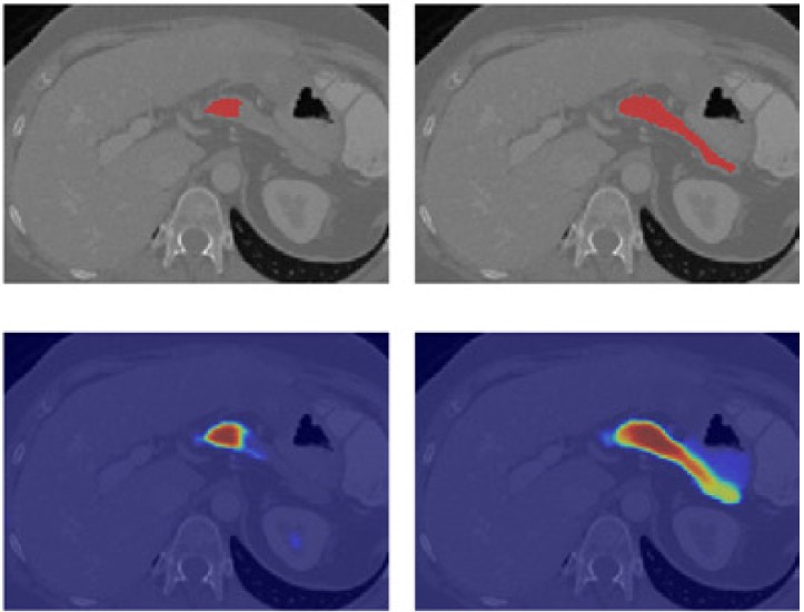
We also highlight how some studies are using advanced techniques like deep learning, segmentation, and radiomics to push the boundaries of accuracy. This review offers a clear picture of how AI can be a game-changer in the diagnosis of liver lesions. Exciting times are ahead for AI in healthcare, and this review brings some of the latest innovations in medical imaging.
Find the link to the paper here: https://link.springer.com/article/10.1007/s00261-024-04597-x


